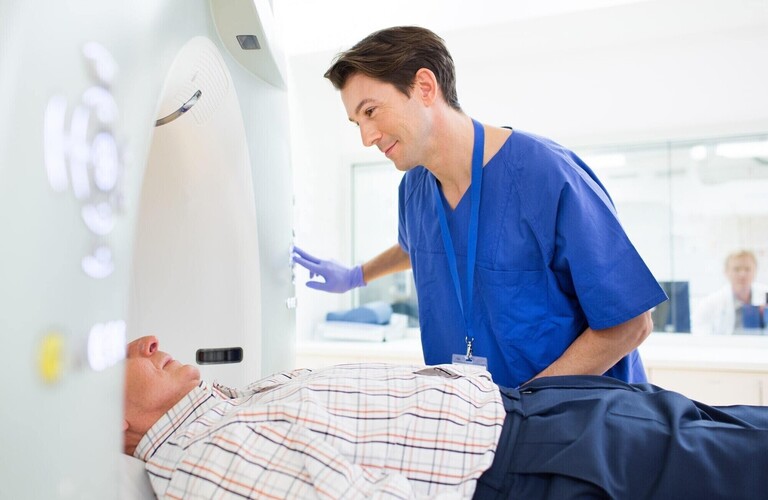
This technique helps the Radiologist and your doctor to understand why you have difficulty passing stool or in some cases why you have inflammation in the lowest part of your bowel.
The scan combines an assessment of the pelvic organs whilst looking for any mechanical reason why you have difficulty opening your bowel.
No preparation is required prior to coming to the centre but after arrival you will have a small enema into the bottom (via a very thin, flexible tube) to help clear the lower bowel. You will then visit the toilet just before the test commences.
For the proctogram, you will be asked to lie on the MRI scanner and a small quantity of special jelly will be introduced into the bottom using a small tube. The tube will then be removed and you will be asked to lie on your back. You may feel worried that this is undignified but please be reassured that we will do our best to ensure your privacy at all times. The procedure is tolerated very well by almost all patients with minimal discomfort.
Examines the pelvis around the anus and lowest part of the bowel for infection or fluid collections. Also frequently used for investigation of patients with anal pain and after surgery.
London Radiology specialises in High Resolution MRI techniques under the supervision of Dr David Burling. This ensures precise diagnosis and assessment of bowel cancer and enables the surgeon and oncologist to determine the best treatment plan. Research on MRI in rectal cancer has shown that careful expert assessment using imaging can help answer the crucially important questions for the patient:
- Can the tumour be removed curatively?
- What type of surgery will be needed?
- Can a permanent stoma be avoided?
- Is preoperative radiotherapy or chemoradiotherapy needed?
- What are the risks of tumour recurring or spreading and how can this be prevented?
- When is the optimum time to undergo surgery after completing radiotherapy?
- Can major surgery be avoided altogether?
The team work closely with the referring surgeons, oncologists and gastroenterologists to ensure comprehensive imaging assessment of the tumour is undertaken before treatments are decided.
A technique used to assess the pelvis after surgery to ensure the surgical join is intact. This will involve a small volume of fluid placed into the bowel via a very thin, flexible catheter inserted carefully into the bottom by an experienced member of the team.
A technique used to examine the small bowel (3 to 4 metres long between stomach and large bowel). This technique involves patients drinking quite a large volume of water (1-2 litres) mixed with a liquid to help the water stay in the bowel (and not be absorbed).
Examines the liver in detail and is often used to complement other imaging techniques such as Body CT. These scans frequently help your Radiologist to characterise a finding on another scan and help work out its significance to you.
A technique used to examine your bile duct system (part of your liver) and is often used to investigate patients with abnormal liver function tests, found on blood tests. If other tests suggest possible bile duct obstruction (most commonly gallstones), MRCP will help identify the cause and site of obstruction.


